
Chinese Fossils shed Light on Single-celled Ancestry of Animals
An international team of scientists have published a remarkable paper detailing their research into an amazing, microscopic fossil that provides evidence of the single-celled ancestor of all complex animal life forms.
The fossil shows an amoeba-like organism dividing in asexual cycles, first to produce two cells, then four, eight, sixteen, thirty-two and so on. The pattern of cell division is very similar to that found in animal embryos, including our own human embryo – but the fossil dates from approximately 570 million years ago, from a geological period known as the Ediacaran (Proterozoic Eon).
The term Proterozoic means “earlier life” in Greek, and this eon covers the time between 2.5 billion years ago up to the beginning of the Cambrian geological period around 542 million years ago. Scientists know that over this immense period of time, life on Earth slowly became more diverse and complex – although all life remained on the microscopic scale up until almost the end of this eon, a time referred to as Neoproterozoic era.
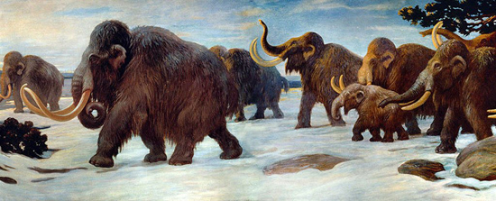
The activity of photosynthetic microbes, transformed our planet providing it with an oxygenated atmosphere, this had first begun in the Archean Eon, but this process continued and in conjunction with climatic changes, simple life forms started to become more abundant. Although, Natural History museums, focus on vertebrate life forms such as dinosaurs and Woolly Mammoths for example, the evolutionary events taking place during the Proterozoic era had a much more significant impact on life on Earth.
During the Proterozoic, cells gradually became larger, more diverse and specialised. Eukaryotes (cells with a nucleus) began to dominate and feeding by ingestion that would eventually lead to the evolution of a gut and digestive system took place for the first time. The transformation of simple cells into these more advanced, specialised cells was probably the longest and hardest step in evolution – demonstrated by the fact that as the Proterozoic gave way to the Phanerozoic (visible life) Eon, there was to be a huge acceleration in evolution – known as the Cambrian explosion.
Fossil evidence of the single-celled ancestors of animals are extremely rare. However, an international team of scientists have discovered such a set of fossils in rocks dating to around 570 million years ago – Middle Ediacaran Period. The fossils were unearthed in southern China. The paper on this discovery has just been published in the journal “Science”. The research team was made up of scientists from the Paul Scherrer Institute, the Chinese Academy of Geological Sciences, Bristol University and the Swedish Museum of Natural History.
Computer Generated Image of Micro-Fossil Undergoing Division

Meet your earliest Ancestor?
Picture credit: University of Bristol Press Release
The computer generated and enhanced image shows 570-million-year-old multi-cellular spore body undergoing vegetative nuclear and cell division (foreground) based on synchrotron x-ray tomographic microscopy of fossils recovered from rocks in South China. The background shows a cut surface through the rock – every grain (about 1 mm diameter) is an exceptionally preserved gooey ball of dividing cells turned to stone.
Single-celled Ancestry
One theory as to the origins of complex life on our planet, proposes that sophisticated eukaryotic cells evolved by the symbiotic fusing of different kinds of bacteria. For example, bacteria capable of fermenting substances merged with swimming, mobile bacteria and the resultant life forms, over hundreds of millions of years merged with oxygenating bacteria and some of these life forms were to become the ancestors of the Animal Kingdom.
However, fossil evidence of these major evolutionary transitions is extremely rare.
The fossils, studied by the international team show in remarkable detail the stages the life cycle of an amoeba-like organism dividing in asexual cycles. Ultimately resulting in hundreds of thousands of spore-like cells that were then released to start the cycle over again. The pattern of cell division is so similar to the early stages of animal (including human) embryology that until now they were thought to represent the embryos of the earliest animals.
Using advanced sophisticated X-ray scanning techniques the scientists were able to view the organisation of cell structures within their protective cell walls. These delicate structures should not have been fossilised but within their marine environment, they became buried in sediments rich in phosphates and it was these phosphates that impregnated the cell membranes, turning them into stone.
Imagine a 570-million-year-old tomb, within which can be found microscopic evidence of cell division.
Lead author on the subsequent research paper Therese Huldtgren, a doctoral student at the Department of Palaeozoology, at the Swedish Museum of Natural History, Stockholm, Sweden commented:
“The fossils are so amazing that even their nuclei have been preserved.”
The powerful X-ray microscopy methodology used by the team revealed that the fossils had features that multi-cellular embryos did not This led the researchers to the conclusion that the fossils were neither animals nor embryos but rather the reproductive spore bodies of single-celled ancestors of animals.
The team used a gigantic microscope in Switzerland to see inside the fossils. This machine is called the Synchrotron, and it is housed in a building the size of a football stadium. It is the one of only a handful of machines of its kind that can produce X-ray images of the magnification and clarity to permit scientists to study micro-fossils in great detail.
Powerful generators fire high-energy electrons around a circular tube, at phenomenal speeds (close to the speed of light). As they travel, they emit X-rays that are so strong that they can penetrate solid rock, and the tiny microscopic fossils, allowing scientists to build up a three-dimensional image of the primitive organism represented by the fossil material.
Professor Philip Donoghue, Professor of Palaeobiology at the School of Earth Sciences, (University of Bristol), stated:
“We were very surprised by our results. We have been convinced for so long that these fossils represented the embryos of the earliest animals, much of what has been written about the fossils for the last ten years is flat wrong. Our colleagues are not going to like the result.”
Professor Stefen Bengtson, Professor of Palaeozoology, at the Swedish Museum of Natural History, Stockholm added:
“These fossils force us to rethink our ideas of how animals learned to make large bodies out of cells.”
Prior to this research such images were interpreted as being the embryos of early animals, sponges or perhaps even the cells of sulphur-oxidising bacteria. Looking at and interpreting the preserved remains of organisms more than 550 million years old which measure just a few microns across is at the cutting edge of palaeobiology, but the team are confident about their findings.
Which Kingdom are You?
")
Animalia, Plantae, Fungi etc.
Image credit: S. A. Museum
The picture above shows another example of a micro-fossil from China. This too dates from the Late Proterozoic Eon. Prior to this new research such fossil images were thought to represent a number of organisms, including embryos of animals that can be found in the Ediacaran fauna, but now some images produced by high-powered X-ray tomography are being interpreted as evidence of the evolutionary origins of multi-cellular life forms.
For models and replicas of prehistoric animals including ancient invertebrates: CollectA Prehistoric Life/Age of Dinosaurs Models.
This is really astonishing. Thanks for highlighting this! -Warren Fahy
As one multi-celled organism to another, you are most welcome.