
CT Scanning an Eagle Lizard
CT Scans Reveal the Armour of Aetosaurs
A student from Bristol University has carried out a study into the armour on the tail of an aetosaur. The study, published in the Scottish Journal of Geology, has provided fresh information on how these large, lumbering herbivores kept themselves safe from ancient predators. Emily Keeble, a recent graduate from the palaeobiology programme at Bristol University carried out the study under the supervision of Professor Mike Benton (School of Earth Sciences). CT (computerised tomography), scans were undertaken, the first time this scientific method has been employed to better understand how the armour of an aetosaur functioned.
A Model of a Typical Aetosaur.
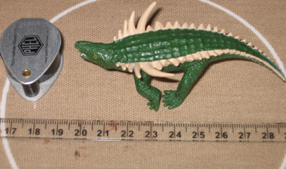
Picture credit: Everything Dinosaur
The picture (above) depicts an aetosaur figure (Desmatosuchus haploceras) from Safari Ltd.
To view the range of Safari Ltd prehistoric animal figures: Safari Ltd. Wild Safari Prehistoric World Models.
Late Triassic Archosaurs
Aetosaurs were heavily armoured, herbivorous archosaurs that were geographically widespread during the Middle and Late Triassic. The term “aetosaur” is from the Greek and it means “eagle lizard”, when scientists first examined the skulls of these animals, their superficial resemblance to the skulls of eagles was remarked upon. The fossil specimens used in this research were collected from a sandstone quarry near the town of Elgin in north-eastern Scotland. They had been donated to the nearby Elgin Museum, where staff members Janet Trythall and Alison Wright were able to identify what they were and arrange for the scanning in Bristol.
Professor Benton explained:
“Aetosaurs were first identified from an Elgin specimen in 1844, but at that time people thought they had found a giant fish. The first specimen showed a number of rectangular scales, arranged in a closely overlapping, regular pattern, and it was called Stagonolepis, meaning drop-shaped scale.”
Dorsal and Lateral Views of the Scottish Aetosaur Stagonolepis
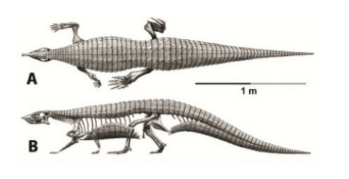
Picture credit: Bristol University (illustration by Jeffrey Martz after work by Alick Walker)
CT Scans Provide Details on Aetosaur Osteoderm Structure
The fossilised tail bones were subjected to high resolution CT scans, this permitted the researchers to see surface details and the texture of the bones and related armour plates.
Emily Keeble added:
“What had been identified as giant fish scales are actually armour plates, or osteoderms, made of plates of bone and embedded in the skin, just like in modern crocodiles.”
Two specimens were studied, both associated with caudal vertebra and possibly from the same animal, but they do not fit together. Each fossil shows a complete circle of osteoderms around the tail, two above, two on each side, and two below.
Caudal Specimen of Stagonolepis robertsoni Used in the Fossil Study
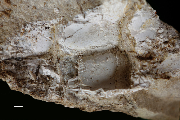
Picture credit: Bristol University
Regular Rows of Osteoderms
The researchers discovered that the rows of osteoderms were very regular and in life covered the entire body from the back of the small head, over the neck, down the back and along the tail. Osteoderms also covered the flanks and underneath. There were even small osteoderms over the fleshy parts of the arms and legs.
The Second Stagonolepis robertsoni Fossil Specimen Used in the Research
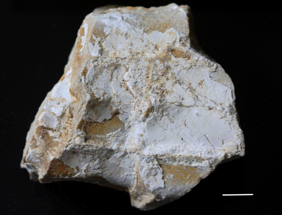
Picture credit: Bristol University
Aetosaur Armour Makes an Effective Defence Against Predators
This study suggests that the armour of these herbivores wrapped around them completely and would have made an effective defence against predators such as rauisuchians and ornithosuchids.
Emily Keeble added:
“Vertebrae of the tail are preserved inside the ring of osteoderms, and these show the specimens were only slightly squished during the preservation process. We could also see how the osteoderms overlap like roof tiles, the osteoderm in front slightly overlapping the one behind. They were linked with connective tissue so the armour overall was flexible, but tough and could probably protect the animal from the fierce predators of its day.”
A Colourised Image of Aetosaur Osteoderms and a Single Scale Shown in More Detail
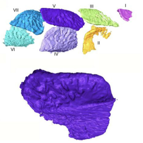
Picture credit: Emily Keeble (Bristol University)
Everything Dinosaur acknowledges the assistance of a press release from Bristol University in the compilation of this article.
The scientific paper: “Three-dimensional tomographic study of dermal armour from the tail of the Triassic aetosaur Stagonolepis robertsoni” by E. Keeble and M. Benton published in the Scottish Journal of Geology.
The Everything Dinosaur website: Everything Dinosaur.