
The Hard, Tough Lives of Ichthyosaurs
A trio of scientists have published a study looking at signs of injury and disease in a range of ichthyosaur genera. Such studies have been undertaken before, indeed the authors of this new paper, published by the Royal Society Open Science, Judith M. Pardo-Pérez, Erin Maxwell (Staatliches Museum für Naturkunde, Stuttgart, Germany) and Benjamin Kear (Uppsala University, Sweden) have examined pathologies in the giant ichthyosaur Temnodontosaurus as recently as 2018, but this study takes a different approach.
The researchers looked in detail at one specific ancient ecosystem, analysing injuries and disease recorded in several different types of ichthyosaur and found some surprising results.
A Scale Drawing Illustrating the Size of the Superpredator Temnodontosaurus

Picture credit: Everything Dinosaur
Ichthyosaur Pathology
Back in 2018, these scientists published a paper detailing the injuries and disease lesions (pathology), associated the ichthyosaur superpredator Temnodontosaurus. They found that despite its size, growing up to ten metres in length, these predators led quite tough lives, given the healed wounds, evidence of trauma and signs of disease preserved in their fossils.
In this new paper, published last week, the scientists examined the fossils of five different ichthyosaurs known from a single fossil deposit (Posidonienschiefer Formation). These fossils from southern Germany, date from the Early Jurassic (Toarcian faunal stage) and represent a marine fauna that suffered a minor extinction event resulting in a significant faunal turnover amongst the vertebrates.
The five genera of ichthyosaur (Posidonienschiefer Formation) from the study in order of maximum size:
1). Hauffiopteryx (2.5 m long) – a relatively short-snouted genus that probably fed on small fish and squid.
2). Stenopterygius (3.5 m long) – feeding on small fish and squid.
3). Suevoleviathan (4 m long)- a primitive member of the Neoichthyosuria clade that with a short-snout that indicates a generalist feeding habit.
4). Eurhinosaurus (7 m long) – its elongated upper snout suggests a specialist position in the food chain, perhaps feeding on small fish or probing the seabed to feed on invertebrates.
5). Temnodontosaurus (up to 10 metres long) – the top predator in the ecosystem, attacking and eating other marine reptiles including ichthyosaurs.
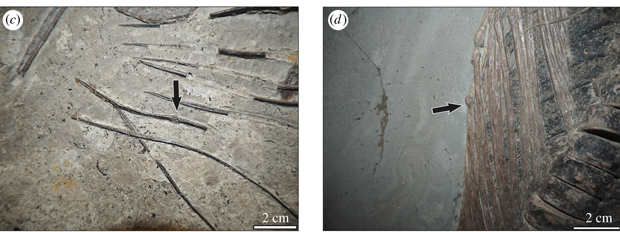
Not Just Damaged Ribs
Damaged ribs are quite commonly found on ichthyosaur fossils, but in this study, a detailed examination of the entire fossilised remains of individual animals was carried out. The team examined the influence of taxa (which species demonstrated the greatest signs of trauma and disease), as well as which parts of the body were damaged the most, the influence of ontogeny and the impact of environmental change (early Toarcian Oceanic Anoxic Event).
Examples of Pathologies in Ichthyosaurs from the Posidonienschiefer Formation

Picture credit: Royal Society Open Science
Small-bodied Genera Do Best
Following the review of the skeletal material, the researchers found that the incidence of pathologies is dependent on the type of taxon being examined. Small-bodied genera such as Stenopterygius had fewer injuries, signs of disease and trauma when compared to larger-bodied ichthyosaurs. Within the Stenopterygius genus, the scientists discovered that more pathologies were identified in large adults when compared to smaller sized individuals. Stratigraphic horizon, a proxy for evidence of change within the ancient marine ecosystem did not influence the incidence of pathology associated with Stenopterygius.
The Research Team Carefully Examined an Extensive Portion of the Posidonienschiefer Formation Ichthyosaur Biota
Picture credit: Royal Society Open Science
Skull and Forelimb Injuries
When all the data from the examined taxa was added together, it was no surprise that the rib area was identified as that part of an ichthyosaur’s body most likely to show signs of pathology. Around 8% of the specimens examined showed rib trauma. However, approximately 6% of skulls and 4% of forelimbs also showed pathologies. In contrast, those areas of the body showing the least signs of injury were the vertebrae and the hind limb.
The researchers concluded that within the fauna studied, ichthyosaurs appear to be similar to living vertebrates in which pathologies accumulate in the oldest/largest members of a population, and larger taxa experience proportionately more frequent skeletal traumas.
The scientific paper: “Palaeoepidemiology in extinct vertebrate populations: factors influencing skeletal health in Jurassic marine reptiles” by Judith M. Pardo-Pérez , Benjamin Kear and Erin E. Maxwell published in Royal Society Open Science.
The Everything Dinosaur website: Everything Dinosaur.

Leave A Comment