
CT Scanner Helps Palaeontologists to Map the Braincase of a Marine Reptile
A farmer’s field in Warwickshire was the site of a remarkable fossil discovery more than sixty years ago. Thanks to the application of advanced medical science and computer modelling, a team of researchers including scientists from Manchester University, have been able to unlock secrets from inside the skull of a giant, Early Jurassic marine reptile. The almost 200 million-year-old fossil, was found at Fell Mill Farm (Warwickshire, England), in 1955. The material included a nearly one-metre-long skull of an ichthyosaur, it had been preserved in three-dimensions permitting scientists a rare glimpse into the internal workings of a prehistoric animal’s skull.
The Beautifully Preserved and Now Fully Restored Skull Specimen
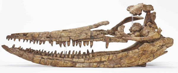
Picture credit: Thinktank, Birmingham Science Museum
Revealing New Information About a Rarely Preserved Marine Reptile Braincase
Most ichthyosaur cranial material is crushed, flattened and distorted during the fossilisation process. This specimen permitted the research team which included Dean Lomax (Manchester University), skilled fossil preparator Nigel Larkin and Laura Porro (University College London), to study a near complete and undistorted three-dimensional skull providing new insights into ichthyosaur cranial anatomy and the morphology of the braincase. Despite the fossil specimen’s excellent preservation, it had never been formally studied prior to this research.
Co-author of the paper, Nigel Larkin explained:
“Initially, the aim of the project was to clean and conserve the skull and partially dismantle it to rebuild it more accurately, ready for redisplay at the Thinktank Museum [Birmingham]. But we soon realised that the individual bones of the skull were exceptionally well preserved in three dimensions, better than in any other ichthyosaur skull we’d seen. Furthermore, that they would respond well to CT scanning, enabling us to capture their shape digitally and to see their internal details. This presented an opportunity that couldn’t be missed.”
Computed Tomography (CT) Scans
To help unlock the information contained inside the skull, the specimen was subjected to computed tomography (CT) scans using a large medical scanner located at the Royal Veterinary College (London). The powerful X-rays in conjunction with computer modelling allowed a three-dimensional and highly accurate digital reconstruction of the fossil to be made. This is the first time a digital reconstruction of a skull and mandible of a large marine reptile has ever been made available for research purposes and to the public.
Going Through the CT Scanner
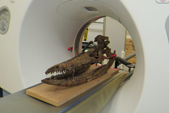
Picture credit: Nigel Larkin photograph taken at Royal Veterinary College, London
Further computed tomography analysis (micro-CT scanning) took place at the University of Cambridge.
Study Clears Up Marine Reptile Fossil Identification
When originally labelled several decades ago, the ichthyosaur was classified as an example of the species Ichthyosaurus communis. Indeed, when Everything Dinosaur wrote an article about this remarkably well-preserved skull back in 2016, the specimen was still being described as Ichthyosaurus. However, lead-author and ichthyosaur expert, Dean Lomax became convinced as the research progressed, that this specimen represented a much rarer species. He identified it as an example of an ichthyosaur called Protoichthyosaurus prostaxalis, the type species of this genus had originally been named in 1979.
To read the 2016 article that describes the skull and shows the post cranial material associated with this specimen: One of Britain’s Largest Ichthyosaurs Goes on Display.
With a skull almost twice as long as any other specimen of Protoichthyosaurus, this is the largest specimen known to science.
Research Team Members View the Results of the CT Scans

Picture credit: Nigel Larkin, taken at the University of Cambridge
Lead-author Dean Lomax stated:
“The first time I saw this specimen I was puzzled by its excellent preservation. Ichthyosaurs of this age (Early Jurassic), are usually ‘pancaked’, meaning that they are squished so that the original structure of the skull is either not preserved or is distorted or damaged. So, to have a skull and portions of the skeleton of an ichthyosaur of this age preserved in three dimensions, and without any surrounding rock obscuring it, is something quite special.”
Protoichthyosaurus prostaxalis
Protoichthyosaurus was first erected by the British palaeontologist Robert Appleby forty years ago. Prior to his research, the fossil material that Dr Appleby assigned to the new genus had been placed in the Ichthyosaurus genus. Indeed, subsequent research challenged this assessment and for some time, the validity of the Protoichthyosaurus genus remained in doubt.
In 2017, Dean Lomax along with colleagues Professor Judy Massare (State University of New York) and Rashmi Mistry (Reading University), conducted a re-examination of the fossil material and carried out extensive comparisons between ichthyosaur and suspected Protoichthyosaurus specimens. The researchers concluded that Protoichthyosaurus was indeed, a valid genus: Reaffirming Protoichthyosaurus as a Valid Genus.
A Life Reconstruction of the Ichthyosaur Protoichthyosaurus prostaxalis
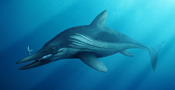
Picture credit: Bob Nicholls @Paleocreations
Back to the Braincase
The skull is not quite complete, but several bones that make up the braincase, which are very rarely preserved in the Ichthyosauridae, are present. The micro-CT scanning conducted at Cambridge University provided crucial data to help reconstruct the internal anatomy of the animal’s skull and brain. The fossil only preserved bones from the left side of the braincase, however, using CT scans these elements were digitally mirrored and 3-D printed at life size to provide a complete braincase.
Commenting on how the use of modern technologies, such as medical scanners, have revolutionised the way in which palaeontologists are able to study and describe fossils, Dr Laura Porro stated:
“CT scanning allows us to look inside fossils – in this case, we could see long canals within the skull bones that originally contained blood vessels and nerves. Scans also revealed the curation history of the specimen since its discovery in the ‘50s. There were several areas reconstructed in plaster and clay, and one bone was so expertly modelled that only the scans revealed part of it was a fake. Finally, there is the potential to digitally reconstruct the skull in 3-D. This is hard (and risky) to do with the original, fragile and very heavy fossil bones; plus, we can now make the 3-D reconstruction freely available to other scientists and for education.”
An Image of the Three-Dimensional Scan of the Protoichthyosaurus Skull Material
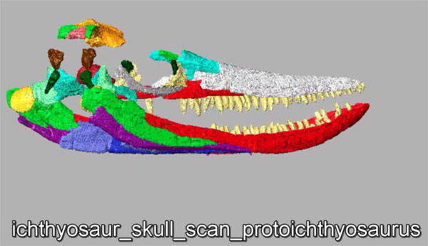
Picture credit: University of Manchester/Thinktank
Dean Lomax added:
“It’s taken more than half a century for this ichthyosaur to be studied and described, but it has been worth the wait. Not only has our study revealed exciting information about the internal anatomy of the skull of this animal, but our findings will aid other palaeontologists in exploring its evolutionary relationship with other ichthyosaurs.”
Visit the Everything Dinosaur website: Everything Dinosaur.

Leave A Comment