
World’s First Named Dinosaur Reveals New Teeth
The fossils that led to the first scientific account of a dinosaur can still provide some surprises, even 193 years after the original paper describing them was published. The first dinosaur to be scientifically described Megalosaurus (M. bucklandii), has stepped once more into the spotlight. A team of researchers have discovered five new teeth within the lower jaw fossil of the world’s first named dinosaur.
Megalosaurus Fossils Used to Describe the First Dinosaur in 1824
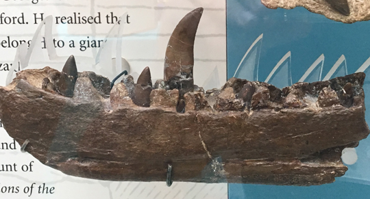
The partial dentary for teeth associated with Megalosaurus bucklandii. Picture credit: Everything Dinosaur.
Picture credit: Everything Dinosaur
The picture above shows the right dentary of Megalosaurus. It is on display at the Oxford University Natural History Museum.
Using state-of-the-art computer tomography scanning technology and three-dimensional computer generated modelling software, the researchers from the Warwick Manufacturing Group (WMG), an academic department at the University of Warwick, in collaboration with scientists from the University of Oxford’s Museum of Natural History have been able to provide new insights about one of the most iconic fossils in the world.
One of the authors of the study, presented this week at the Institute of Electrical and Electronics Engineers (IEEE)’s conference in Italy, Professor Mark Williams stated:
“Being able to use state-of-the-art technology, normally reserved for aerospace and automotive engineering, to scan such a rare and iconic natural history specimen was a fantastic opportunity. When I was growing up I was fascinated with dinosaurs and clearly remember seeing pictures of the Megalosaurus jaw in books that I read. Having access to and scanning the real thing was an incredible experience.”
Famous Dinosaur Jawbone
In 1824, the Reverend William Buckland published a description of various fossils that had been found as quarrying tunnels were excavated at Stonesfield, north of Witney in Oxfordshire. The fossils had been found some years before, the dentary having been placed in the collection of the Oxford Anatomy School at Christchurch College (Oxford) in 1797. Reverend Buckland believed the fossilised bones and teeth came from a giant, antediluvian lizard, hence the name “Big Lizard”, Megalosaurus having been proposed by James Parkinson in 1822.
A 19th Century Interpretation of Megalosaurus Compared to a Modern Interpretation of M. bucklandii
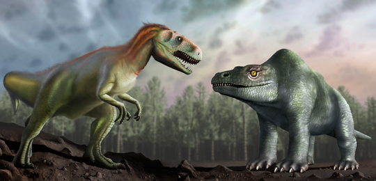
A modern interpretation of Megalosaurus (left) with a reconstruction based on the original illustration by Richard Owen (right).
Picture credit: University of Warwick/Mark Garlick
The illustration above shows an artist’s impression of how Victorian palaeontologists such as Richard Owen thought the Megalosaurus looked (right), compared with a modern interpretation of this Middle Jurassic carnivore.
Digital Three-dimensional Image of the Dentary
Using state of the art CT scanning technology and specialist three-dimensional analysis software, Professor Williams took more than 3,000 X-ray images of the world-famous Megalosaurus jawbone, creating a digital three-dimensional computer generated image. The image revealed five previously unseen teeth embedded in the dentary and also provided important insights into historical repairs. It turns out that there is actually less plaster and filler in the fossil, as this technique has allowed scientists to see the extent of the infilling and repairs for the first time.
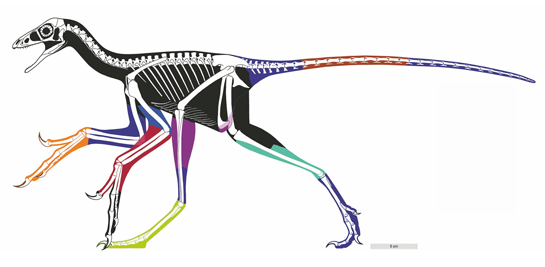
Megalosaurus bucklandii was Probably an Apex Predator
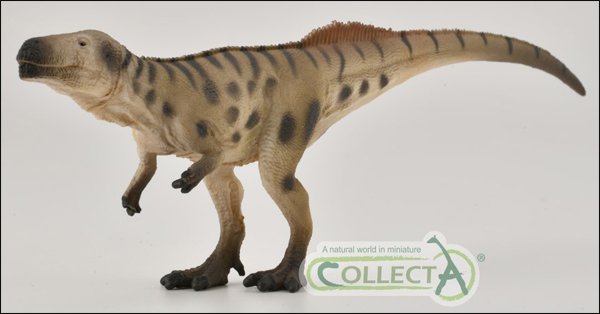
The new for 2021 CollectA Megalosaurus in ambush dinosaur model.
The picture (above) shows the CollectA Megalosaurus in ambush dinosaur figure.
To view this range: CollectA Age of Dinosaurs Popular Figures.
Megalosaurus Fossil Jawbone
The specimen is damaged, it is likely that some of the damage occurred when the fossil was excavated but over the two hundred years since the fossil was found some restoration work has taken place. For example, records at the Oxford University Museum of Natural History, where the specimen is housed, show that sometime between 1927 and 1931 repairs to the jawbone took place. The scans show the true extent of repairs on the fossil for the first time, revealing that there may have been at least two phases of repair, using different types of plaster. This new information will help the museum make important decisions about any future restoration work on this iconic fossil.
The analysis also revealed the presence of five teeth that had not been detected before. The teeth consist of the remains of old, worn and broken teeth plus embryonic replacement teeth. Unlike us, Megalosaurus was able to continually replace its teeth throughout its life. The replacement tooth grew inside the jaw, adjacent to the root of the active tooth on the lingual* side of the jaw.
A full-sized, but very thin crown formed first and this grew in thickness as more layers of dentine were added. The growth of the embryonic tooth placed pressure on the active tooth root, causing the root to become slowly reabsorbed into the jawbone. The replacement tooth was able to push itself inside the old tooth root and effectively usurp that tooth from the socket in the jaw where it had been located. The old, worn tooth having been weakened, would most likely break and the crown would be lost, permitting the younger tooth to replace it in the jawline. A similar process is seen in extant Crocodylia today.
Helping to Identify Forgeries
This research was made possible through a collaboration between Professor Williams’ research group at WMG, University of Warwick – including PhD researcher Paul Wilson – and Professor Paul Smith, director of the Oxford University Museum of Natural History. When not being scanned or used in other research, the Megalosaurus jawbone forms part of an extensive British dinosaur fossil display at the Oxford University Museum of Natural History.
An ability to utilise a non-invasive technique to map fossil material provides palaeontologists and conservators with vital information about the preservation status of a specimen. It also identifies and maps any repairs that have taken place previously. In addition, this technique which does not harm the fossil, can detect the presence of filler and other modifications often added by unscrupulous dealers to raise the potential value of their fossil finds.
Forgeries and hoaxes have no hiding place when it comes to CT scans.
The research was recently presented at the Institute of Electrical and Electronics Engineers (IEEE)’s International Instrumentation and Measurement Technology Conference in Torino, Italy.
The scientific paper, “Utilising X-Ray Computed Tomography for Heritage Conservation: The case of Megalosaurus bucklandii”
Visit the Everything Dinosaur website: Everything Dinosaur.


Leave A Comment